Trial Case and Lenses


Trial Case and Lenses

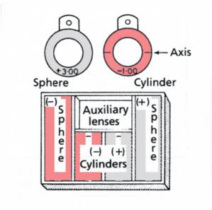
The trial case is a tray of lenses and accessories used to determine the refractive error of an eye. These lenses are individually marked in the strengths of dioptric power of each lens, as well as in the direction of axis of the cylindric lenses. The trial case consists of:
• a pair of plus spheres ranging from +0.12 to +20.00 D
• a pair of minus spheres ranging from -0.12 to -20.00 D
• a pair of plus cylinders ranging from +0.12 to +8.00 D
• a pair of minus cylinders ranging from -0.12 to -8.00 D
• accessory lenses
• trial frame.

These lenses are designed to fit a standard trial frame. Each lens is encircled by a metal rim for protection.
Handles are provided with spheres for ease of handling and are optional with cylinders. The choice is governed by the type of trial frames used. The cylinder is marked with reference to its axis and not the meridian.

Accessory lenses available in a trial case are:
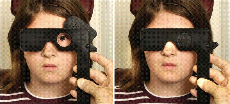
• an occluder lens
• a pinhole disk
• a stenopeic slit
• a Maddox rod lens
• prisms ranging from 0.50 to 6.00 prism D
• a red glass filter lens.
• a pair plano lenses

Faculty of Optometry and Visual Sciences in Sudan


Faculty of Optometry and Visual Sciences

Faculty of optometry and visual sciences, Alneelain University in Khartoum – Sudan.
Established in 1954 as institute of optometry in Khartoum eye hospital. Joined ministry of Higher Education in 1986 as the High Institute of Optometry, and lastly was annexed to Alneelain University in 1997 when it was re- named to become Faculty of Optometry and Visual Sciences (FOVS).
Currently FOVS has the following programs:
1- BSc optometry in 5 years with sub- specialization in either orthoptics, contact lenses, ocular photography or ocular neurology.
2- Diploma in ophthalmic technology in 3 years.
3- Diploma in optical dispensary in 3 years.
FOVS also offers MSc and PhD degrees in Optometry.
FOVS is the only of its kind in Sudan and is the first in Middle East and Africa. There is similar colleges in only 30 out of 193 countries in the world.
In 2010, Alneelain University Eye Hospital was established as part of FOVS to expand training capacity and to serve community.

This entry was posted on February 14, 2014. 2 Comments

How to create a prismatic prescription through decentration!


1

Normally, eyeglasses are fitted with the optical center of the lens directly in front of the eye. If the lens is fit off-center, image displacement can occur due to induced prism. The higher the power or the further the lens is fit off-center, the higher is the induced prismatic effect.
Prentice’s Rule:
Prism Power = Lens Power x Decentration (in centimeters)

Induced Prism
To better understand why there is induced prism, the cross-section of a plus lens can be likened to two prisms base-to-base, as the lens is thicker in the middle and thinner at the edges. Likewise, a minus lens can be likened to two prisms apex-to-apex, thinner in the middle and thicker at the edges. This induced prism can actually be used to the advantage of the lab when prism is called for in a prescription. This is known as prism by decentration.
Decentration = Prism * 10 / Power
This gives you the decentration in mm.

If (power0)
Base In Prism, decentration = –
Base Out Prism, decentration = +
Base Up Prism, decentration = –
Base Down Prism, decentration = +

Example:

-5.00sph lens need with 2.5∆ base in
Decentration = 2.5 x 10 / -5 = 5mm
As this is base In prism and the power is minus(<0), so that you have to add 5mm to the mono PD to get the required prism(2.5∆).

Note:

Important thing to remember: Prentice’s Rule breaks down for ophthalmic lens powers of less than about ± 1.00diopter.

Pin Hole Test


Image

Pinhole test is an effective tool to determine if distance visual acuity can be corrected with glasses or contact lenses.

            The pinhole disk admits only central rays of light, that do not need to be refracted by the cornea or lens to fall to a point on the retina as opposed to a circle of blur. A single pinhole of not more than 2.4mm or a multiple circular arrangement of 1.0mm pinholes can be used.

            Upon reevaluation with the pinhole, if a patient’s visual acuity improves two or more lines, there is probably a refractive error present, and refraction (evaluation for corrective lenses) should be performed before any further testing.

             If the acuity is not improved with pinhole evaluation, it is likely that the cause of the decreased visual acuity is not refractive, and further ophthalmologic evaluation is indicated.

            Performance of the Pinhole Acuity should include the following steps:

  1. Position the patient with corrective lenses.
  2. Occlude the eye not being evaluated and position the pinhole(s) in front of the eye being evaluated.
  3. Instruct the patient to look through the pinhole(s) at the distance chart.
  4. Instruct the patient to use small movements of the paddle to align the pinhole(s) so they can read the optotypes from right to left.
  5. Have the patient begin reading on the last line that was seen without the pinhole(s)
  6. The smallest line read from the distance Snellen chart is recorded in the patient’s medical record following the already recorded distance acuity.
  7. Repeat above procedure for the fellow eye.

Visual Acuity Test


Chart1

Measurement of visual acuity is necessary to determine the integrity of the visual system. When performed as part of the comprehensive ocular examination, visual acuity assessment can reveal refractive errors, optical disorders, and ocular disease.

Visual acuity should be evaluated both uncorrected and corrected, at distance and near, with each eye being tested independently. Binocular acuity is indicated in specific situations such as nystagmus (repetitive involuntary eye movements).

The distance acuity is typically measured at twenty feet (six meters) and most optotype charts have been calibrated to this standard. At twenty feet the rays of light from a distant object are practically parallel, and little accommodation is required. If the examination lane is shorter than twenty feet, mirrors and a reflective system are used to achieve the appropriate test distance. In some instances an optotype chart may be recalibrated for shorter testing distances.

What is Optometrist?


IMG_2735

Optometrists are independent, primary health care providers who examine, diagnose, treat, and manage diseases and disorders of the visual system, the eye and associated structures as well as diagnose related systemic conditions.

Optometrists are an integral part of the health care team. As eye care practitioners, they are skilled in the co-management of eye health and vision care. They examine the structures of the eye to detect and diagnose:
•Vision conditions such as nearsightedness, farsightedness, astigmatism and presbyopia
•Binocular vision conditions such as convergence insufficiency, which can cause eye discomfort and difficulty reading
•Eye diseases such as glaucoma, cataracts and retinal disorders
•Systemic diseases such as hypertension and diabetes

Optometrists prescribe and/or provide eyeglasses, contact lenses, low vision aids and vision therapy. They prescribe medications to treat eye diseases and perform certain surgical procedures. Optometrists also do testing to determine the patient’s ability to focus and coordinate the eye, judge depth perception, and see colors accurately.